2015 preamble: Hello dear site visitor. Welcome to the site.
You have chosen to check out a 2015 posting. 2015 was my breakthrough year. Up till then I’d been wedded to direct scorching of hot metal template (whether fully 3D or bas relief). Why? The resulting imprints were not unlike TS body image, albeit sharper, but were, nevertheless, negative tone-reversed, responsive to 3D-rendering software (ImageJ) , bleachable etc etc. (All very encouraging).
But there was a fly-in-the-ointment: one cannot monitor degree of coloration when metal is pressed into linen – the action/contact zone being shielded from sight.
The solution took shape in late 2014: instead of heating the template, I coated it with a thin smear of oil, then lightly dusted with white flour ,then imprinted the flour-image onto linen. I then heated the linen over a ceramic hot plate (later with a modern electric iron or fan oven). Result: an arguably better model of the TS body image (fuzzier etc)
But the full potential of the was new procedure not realized. Why? Because I imprinted onto dry linen.
Similar two-step technology was developed in 2015 via an entirely different route.
I had been looking at acids as possible colorants for creating a look-alike “Shroud” image (with most or all its distinctive and unusual features) . Weak organic acids and even strong mineral acids (sulphuric etc) were quickly rejected for one reason or another. They either failed to produce colour, or, if they did (sulphuric) it was not only scarcely visible, but the cloth was dramatically weakened.
It was nitric acid (HNO3) that proved interesting. It produced a faint yellow coloration of untreated linen, probably on account of protein traces.. Could that be made more pronounced? Answer: YES, first by coating the linen with a high concentration of protein (gelatin or egg white), then realizing that a more common medieval commodity, i.e. plain white flour with its 10% or thereabouts of protein would serve.
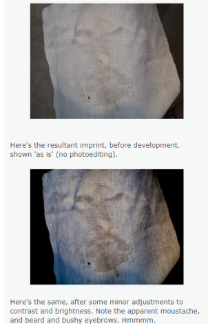
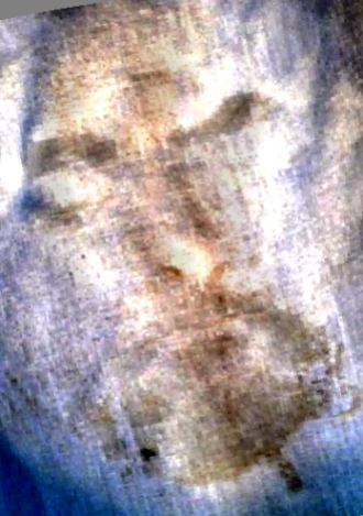
Image formation? Initially I created imprints of metal and other templates (ceramic etc) using a slurry of wheat flour in water, with or without heat treatment to gelatinize the starch. I found to my delight that I could produce a negative image of my face, 3D enhancible, simply by taking the flour imprint and bolding up the intrinsic faint yellow colour of wheat flour using simple commercial photoediting software (Windows Office).
Insert image of my face, from May 2015: get it from this link.
Link:
http://colinb-sciencebuzz.blogspot.com/2015/05/modelling-turin-shroud-my-flournitric.html
Images of hands etc, brass crucifixes, plastic figurines quickly followed, still using liquid flour slurry as imprinting medium.
But they were criticized by a particular high-profile individual: I was told the edges were too sharp, too well defined to be considered a valid model.
There was a simple solution that largely silenced the critic.
I returned to the Oct 14 technique that had been shelved, with a small modification: smear the hand, (or metal or plastic template) with veg oil, sprinkle with white flour, shake off excess flour, imprint onto WET linen, then heat the linen until the desired degree of yellow coloration had been obtained (via the kind of Maillard browning or caramelization reactions that occur rotinely when baking flour-based goods).
Yes, nitric acid was dispensed with as the means of coloration. In its place was thermal colour development. Expressed more simply: HEAT the imprinted linen!
No, not a scorch, in the sense that the linen constituents per se (cellulose etc) were being coloured via complex thermal reactions, but, totally separate, at least chemically the acquired flour imprint. (How that imprint interacts physically with linen fibres provides a whole new dimension for speculation and detailed research – with microscopy leading the way.
One final step: a vigorous rub with soap and water to dislodge loose encrustations. What stubbornly remains behind is a somewhat diffuse fuzzy-edged yellow to brown discoloration of the cloth. If imprinted as an image, one sees with time and further reseacrh (2015 and beyond) a growing list of resemblances to (guess what?) the reported properties of the TS body image, including those bizarre features revealed by microscopy (“half tone effect”, “image discontinuities” etc). Yup, I say we’re home and dry with “Model 10”. Correction – home and wet (after the final washing step). Just add 30 mins for drying and we’re home and dry!
It did not take long for the rest of the faux-biblical ‘narrative’ pieces to fall into place. A fuzzy whole body imprint obtained with white flour and heating, with blood in all the right places, is likely to have been a 14th century Veil of Veronica -obsesssed attempt to simulate (“fake”) the imprint that might have been left on Joseph of Arimathea’s fine linen, used to retrieve a recently crucified body from a cross, for transport to a nearby rock tomb. (NO, neither intended nor used as the final burial shroud in 33AD, taking the biblical account as, er, 100% Gospel truth, thereby making the term “Turin Shroud” highly misleading, especially as it’s routinely expanded to “burial shroud”). See margin comments on this site for more details.
Here are the two key links: the first to the floured-up horsebrass from 2014:
http://colinb-sciencebuzz.blogspot.com/2014/10/modelling-shroud-of-turin-image-with.html
The second is the link to imprinting of my face with plain white flour (amazingly) WITHOUT use of physical developing agent (mere recourse to photoediting software):
http://colinb-sciencebuzz.blogspot.com/2015/05/modelling-turin-shroud-my-flournitric.html
Maybe three more links from red-letter year, 2015, like Dan Porter’s reports on my Model 10 thinking, proposed mechanism for “half tone effect etc etc.
So much for the background to 2015. (Thanking you dear reader in advance for your patience and forebearance. (Hopefully you’ll appreciate that when one has posted a reported-in-real time online learning curve via 350+ postings, here and elsewhere, some kind of packaging then makes sense).
Here’s the start of the original posting.
OK, so the 1981 STURP summary did not describe the enigmatic image of the Man on the Turin Shroud as a “scorch”. The term “scorch” appeared nowhere, and this investigator no longer uses that term anyway, for reasons that will shortly be explained.
But the STURP summary came PDC to describing the image as having the characteristics of a scorch, as per the brown coloration one sees on linen when a hot iron on maximum setting (“linen”!) is held against it for too long. I quote (my bolding):
Furthermore, experiments in physics and chemistry with old linen have failed to reproduce adequately the phenomenon presented by the Shroud of Turin. The scientific consensus is that the image was produced by something which resulted in oxidation, dehydration and conjugation of the polysaccharide structure of the microfibrils of the linen itself. Such changes can be duplicated in the laboratory by certain chemical and physical processes. A similar type of change in linen can be obtained by sulfuric acid or heat.
Note the reference to “heat” (or acid) in the final sentence that is suggestive of a scorch mechanism, or at any rate “thermally-assisted” imprinting, such that the linen carbohydrates lose water molecules (“dehydration”), then undergoing further chemical change (“oxidation”, “conjugation” ) that results in the tan or sepia coloration.
Sure, that seemingly specific reaction mechanism, based on re-arranging chemical structure with heat (or acid) as the input energy then gets clouded by what follows:
However, there are no chemical or physical methods known which can account for the totality of the image, nor can any combination of physical, chemical, biological or medical circumstances explain the image adequately.
Thus, the answer to the question of how the image was produced or what produced the image remains, now, as it has in the past, a mystery.
But it was a start, for this investigator at any rate, one who has explored both heat and acid as a means of imprinting tan-coloured images on linen (links can wait). The first approach involved heating up bas relief templates (horse brasses etc) then pressing onto linen, and later still full 3D templates (e.g. brass crucifix) and obtaining what he regarded as promising results i.e. negative 3D-enhancible images, albeit with unanswered questions re superficiality still to be explored, both by me AND by sindonology generally, pending a breakthrough in image-probe techniques).

Old (Mark 1) direct scorch technology, now superceded by the two-stage Mark 2 flour imprinting technology (see banner). It’s inserted here merely to show how well a scorch imprint responds to 3D enhancement, before and after performing a Secondo Pia style reversal of light v dark on the initial negative imprint. (Yes, it is imprinting rather than painting that explains why the TS body image is a negative, the latter not being exclusive to photography as so many seem to imagine).
Later, that model was superceded by the Mark 2 system, in which hot metal templates were abandoned. In their place was imprinting onto linen from real body anatomy (yipee, my own hand!) using dry white flour onto dampened linen, and then roasting the imprinted linen in a hot oven to produce a sepia image (Mark 2 model). But Mark 1 and Mark 2 could both be described as thermal imprinting mechanisms, even if the term “scorch” model was appropriate for the first only.
But as soon as the Mark 1 model was proposed with its hot metal template, this investigator immediately attracted hostile targeted flak, from Mr.Big of sindonology, a surviving STURP team member no less. Here’s what he (STURP’s Documenting Photographer) said in a comment to the shroudstory site in Feb 2012 that now, nearly 4 years later, I shall shortly be able cautiously to respond to experimentally in three short postings, of which this one is the first.

Note the assumption that the scorch marks on the TS acquired in the 1532 fire are an appropriate benchmark for heat imprints generally, despite the precise conditions (temperature, oxygen, alleged contact with molten silver(?) etc being unknown. How scientific is that?
To business: here’s the uv lamp being put through its paces. The label on the box reads “Mains Powered UV Bank Note Checker”, so that was the first test – to see if it showed up something on a bank note normally not visible.
Yes. It is fit for stated purpose – showing up the fluorescent security feature on a banknote.
Would it work with fluorescent marker pens?
Answer – yes. Spectacularly so.
And here’s a wider range of fluorescent marker pen inks, under the uv lamp.
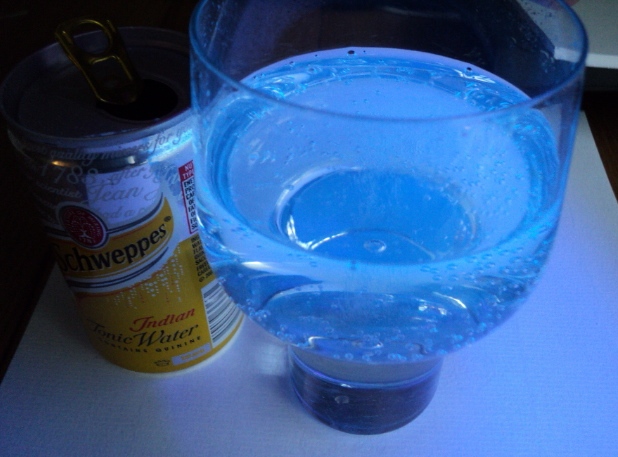
Tonic water is famous for its blue fluorescence, sometimes visible in bright sunlight for those who like a G/T on the patio. It’s due to the quinine that gives tonic its bitterness. Here it is under the uv lamp:
Now for a more demanding test. Riboflavin, aka Vitamin B2, is well known for its yellow-green fluorescence. Was there a source in the house, without going out to buy vitamin pills? Possibly, given there’s some Marmite in the house – “you either love it or loathe it” – (autolysed yeast extract), rich in B vitamins.
A little of the thick brown spread was dispersed in cold water to give a pale brown coloration, some of which was then spotted onto white tissue.

Testing marmite solution under the uv lamp. That’s the brown stuff in the middle with a faint yellow-green halo under uv.
Problem: whilst one could see a faint green fluorescence under the uv lamp, especially when held close to the source of ‘black light’, the fluorescence was scarcely visible in the photograph, appearing bluish-white rather than green. We had reached the operating limit of the uv lamp, designed for less demanding tasks like detecting forged banknotes.
What about glass? Does it filter out uv, as frequently stated or assumed? I see no evidence for that, at least with the uv from the present source.
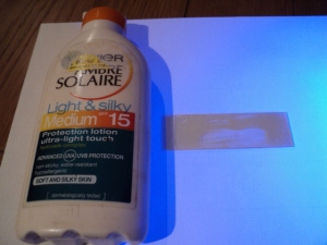
What about sunblock, aka sunscreen?

Sunblock smeared onto glass slide, second slide then placed on top to make a thin sandwich. Viewed first in ordinary visible light -scarcely visible.
Here’s the same after switching on the uv lamp (the latter right, out of picture):

As above, illuminated with uv. Note the way the sunblock does what it says on the label (except for thinner smear lower centre).
Let’s test the smeared slides as a rough-and-ready uv filter, given that glass alone does not work:
So what can I say, scientifically speaking, about the composition of the ultraviolet radiation from my recent high-street purchase (major electrical retailer, Strand branch, London, UK). Answer: nothing. The unit came without a word of description on the box, apart from “Mains Powered UV Bank Note Checker, DETECTEUR DE FAUX BILLETS, UV Lamp shows Hidden Ultraviolet Features, Suitable for Most Currencies, Compact Design is Ideal for all Locations, Rugged Black Plastic Case, Mains Powered. Colour…Black; Power(W)…4; Dimensions(mm)…75x120x180;Weight (kg) …0.36
Brand name? None shown on the unit itself (it says EAGLE on the box, though that may be the importer/distributor, for which an address is given on Merseyside.
Country of origin? Nope, we’re not even told that, though one might make an educated guess.
There’s not even a booklet or so much as a leaflet inside the box, summarising technical details. What’s providing the uv radiation? It’s a cylindrical glass tube that glows violet. I suspect it contains mercury vapour, and provides mainly longwave ultraviolet, probably in the range 315nm – 400nm, aka UVA. But that’s a pure guess, maybe an educated one (I spent 2 years in the States in the early 70s irradiating bilirubin with intense white light with a strong blue component, but quickly dispensed with ordinary fluorescent tubes. Instead, I took the thick shield off of a high pressure mercury lamp designed to provide filtered uv, and simply had to accept that any results I obtained might have been due to the minor uv rather than major visible light component).
Is my lamp suitable for a scientific re-investigation of uv fluorescence, as it relates to the Turin Shroud, especially the STURP reports re the body image being non-fluorescent, compared with the 1532 burn holes that were?
Answer: No, definitely not. I am not here to posture as if still a working scientist (I’ve been retired since 2002).
So why am I writing this post, the first of three?
Answer: because I now see my role more as a crime detective, looking for and sifting clues, seeking to ascertain who is telling the unadorned truth, and who ain’t, and indeed is attempting to bullsh*t with pseudoscience (and have already made significant progress seen in those brutally simple terms).
So don’t expect to see long technical screeds, table of numerical data etc etc. The most one can expect is to see my finger pointing in a certain direction, saying “that’s the way to go if you want my honest and candid opinion”. Maybe you don’t, in which case you either aren’t reading this post, or, if you are, may decide not to read the next two, based on findings with my high street bank note checker.
From the internet:
UVR (UV radiation) is divided into wavelength ranges identified as UVA (315 to 400 nm), UVB (280 to 315 nm), and UVC (100 to 280 nm). Of the solar UV energy reaching the equator, 95% is UVA and 5% is UVB
From this site: Sunscreens explained:
Sunscreens are products combining several ingredients that help prevent the sun’s ultraviolet (UV) radiation from reaching the skin. Two types of ultraviolet radiation, UVA and UVB , damage the skin, age it prematurely, and increase your risk of skin cancer.
UVB is the chief culprit behind sunburn, while UVA rays, which penetrate the skin more deeply, are associated with wrinkling, leathering, sagging, and other light-induced effects of aging (photoaging ). They also exacerbate the carcinogenic effects of UVB rays, and increasingly are being seen as a cause of skin cancer on their own. Sunscreens vary in their ability to protect against UVA and UVB.
Posting 2 in this series will compare the effect of uv light from the new lamp on (a) Mark 1 contact scorches (hot metal template) with the margins of full-thickness burn holes in linen, the latter modelling the 1532 Chambery fire.
Posting 3 will look at the Mark 2 imprints obtained with white flour and a hot oven.
Time scale? Maybe a week, at most two.
Colin Berry
December 8, 2015.


