Late addition (July 2019)
Please forgive this postscript, correction, “prescript”, correction, intrusion, added many years later – based on some 350 and more postings here and elsewhere.
That’s including some 7 years of my hands-on investigation into image-forming techniques, chosen to be credible with simple, indeed crude, medieval (14th century) technology etc etc.
(Oh, and yes, I accept the radiocarbon dating, despite it being restricted to a single non-random corner sample, making all the oh-so-dismissive, oh-so-derogatory statistics-based sniping totally irrelevant – a ranging shot being just that me dears- a single ranging shot, albeit subdivided into three for Arizona, Oxford and Zurich).
Sindonology (i.e. the “science” , read pseudoscience – of the so-called “Shroud ” of Turin) can be simply summed up. It’s a re-branding exercise, one designed to pretend that the prized Turin possession is not just J of A’s “fine linen”, described in the biblical account as used to transport a crucified body from cross to tomb.
Oh no, it goes further, much further, way way beyond the biblical account. How? By making out that it was the SAME linen as that described in the Gospel of John, deployed as final “burial clothes”. Thus the description “Shroud” for the Turin Linen, usually with the addition “burial shroud”. Why the elision of two different linens, deployed for entirely different purposes (transport first, then final interment)?
Go figure! Key words to consider are: authentic relic v manufactured medieval icon; mystique, peaceful death-repose, unlimited opportunity for proposing new and ever more improbable image-formation mechanisms etc. How much easier it is to attach the label “Holy” to Shroud if seen as final burial clothes, in final at-peace repose – prior to Resurrection- as distinct from a means of temporary swaying side-to-side transport in an improvised makeshift stretcher !
As I say, a rebranding exercise (transport to final burial shroud) and a very smart and subtle one at that . Not for nothing did that angry local Bishop of Troyes suddenly refer to a “sleight of hand” after allegedly accepting it when first displayed. Seems the script was altered, or as some might say, tampered with! It might also explain why there were two Lirey badges, not just one. Entire books could be written on which of the two came first… I think I know which, with its allusion (?) to the Veil of Veronica… yes, there are alternative views (the face above “SUAIRE” a visual link to the face-only display of the Linen as the “Image of Edessa” or as that on the then current “Shroud” per se.

Face shown (left) on mid- 14th century Machy Mould (recently discovered variant of the Lirey Pilgrim Badge) above the word “SUAIRE” (allegedly meaning “shroud”). Inset image on the right: one version among many of the fabled “Veil of Veronica” image. I say the two are related, and deliberately so, but this is not the time or place to go into detail.
No, NOT a resurrectional selfie, but instead a full size version of, wait for it, the legendary VEIL OF VERONICA , product of inital body contact – no air gaps- between body and fabric, but with one important difference. The Turin image was intended to look more realistic, less artistic.
How? By displaying a negative tone-reversed image implying IMPRINT (unless, that is, you’re a modern day sindonologist, in which case ‘resurrectional proto-photographic selfie” becomes the preferred, nay, vigorously proferred explanation assisted by unrestrained imagination, creation of endless pseudoscience etc etc, with resort to laser beams, corona discharges, nuclear physics, elementary particles, earthquakes etc etc – the list is seemingly endless!
Welcome to modern day sindonology.
Personally, I prefer no-nonsense feet-on-the-ground hypothesis-testing science, aided by lashings of, wait for it, plain down-to-earth common sense.
Start of original posting:
Mario Latendresse’s Shroud Scope images are fine as far as they go. They retain high definition, aka high-resolution (HD and HR respectively) up to to their top level of magnification.

Epsilon-shaped bloodstain, forehead, Man in the Shroud (from Mario Latendresse’s Shroud Scope archive, top magnification, contrast/brightness enhanced)
But they lose resolution and become pixellated if one tries to magnify further.
Why would one want to magnify further? Answer: to investigate the region between seeing the herringbone weave, with blood image apparently confined to the ribs of the weave, and the individual interwoven fibres. There is a paucity of such images available on the internet – they appear to be in the Mark Evans’ archive held by Barrie Schwortz and his STERA Inc. as recommended to me recently by Dr.Thibault Heimburger. Example:
One needs to know which of the latter are contributing to the herringbone weave, and which are not, and the only way of doing that reliably is to zoom in and zoom out on a single image with an unbroken continuum until one has a feel for the transition between photomacrograph and photomicrograph respectively.
———————————————————————————-
(Note added 14 April 2013: when I wrote this last July, I had not appreciated that the ribs of the herringbone weave ARE discernible in the above picture. They are the stacked but staggered horizontals (running diagonally from top left to bottom right) each of which is 1 thread passing over 3, before going under the next, then over the next 3 etc. I’m deliberately avoiding all mention of warp and weft here, given the later controversy as to which is which, and the lack of what one might describe as an unambiguous scientific definition that allows one to be absolutely certain as to which is which without being present at the loom during weaving).
———————————————————————————-
Barrie Schwortz may have the images in his copyright-protected archive, but has stated (my bold) :
“Due to copyright considerations, we cannot provide high resolution digital files without a written licensing agreement. We do not permit our high resolution files to be published on the internet.”
Why not Mr.Schwortz? Correct me if I’m mistaken, but I thought your STERA existed to promote Education and Research…
It might be more accurate to describe STERA as a copyright organization that exists to promote STERA, as this link should prove beyond any shadow of doubt. (Why should STERA hold the copyright on the iconic Shroud photographs taken by Secondo Pia – to take just one example from a long long list).
Update 13th April 2013 : we now have the Shroud 2.0 App with higher resolutions than we have seen previously, at least for those with access to a smartphone.
I’ve been playing around a bit with the image that appeared on the Daniel R.Porter’s shroud.com site, showing a close-up of the epsilon (reversed 3) bloodstain on the forehead.
Here’s the image as shown, with the blood just visible on the 1-over-3 threads comprising the ribs of the herringbone weave (quite how much is in those furrows is anyone’s guess, but I suspect it is smaller than one would think at first sight)
Note that the pink colour has intensified somewhat on the 1-over-3 threads, and there is a hint of a more intense yellow in places outside the blood stain corresponding with faint patches of body image.
Note further yellowing /intensification of the body image colour, but the yellow is now replacing pink in the bloodstain too.
Note how the initial pink coloration of the threads in the epsilon has now been replaced by the yellow coloration of body image at high contrast. Artefact? Or is there body image under the epsilon bloodstains that has been unmasked by increasing the contrast? More to follow (control experiments).
Futher update: here, in response to an enquiry, is the frontal and reverse side of some fabric that has been dabbed with blood.
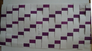
Late addition (July 1 2013): herringbone weave modelled using a suggestion from Hugh Farey on the James Randi Forum (more details to follow). I chose this old post as one having the greatest relevance to the matter of that (enigmatic) herringbone weave.

Underside – the purple strip is still the crown thread, as per topside, but now accounts for 25% of the total (cf 75% on top side)
Late edit (8 years later – Aug 18, 2020)
I realized just a few days ago that my modelling above was incorrect. How did I know? Answer: I came across a STERA-owned photograph of one of the portions of the TS taken for radiocarbon dating (probably Arizona’s). They didn’t match. So I’ve spent half the morning with coloured paper and guillotine, and come up with the following which, hooray, does match! (The error is to do with the manner in which the 3/1 “herringbone weave” is staggered – nuff said…)

I shan’t bother displaying the reverse-side image. It generally causes much head-scratching (my own included!).


I agree 100 percent Colin. Finding a decent resolution image of the shroud is so hard to come by. It’s down to Turin in my personal opinion. They dont want other people making money on what they say is theres. The shroud should not belong to any 1 person in my opinion. It’s either that or there are things on the HD images that they dont want researchers finding out lol!. What images do you use personally? Maybe we could do a swap? I have Durantes 2000 and 20002 images (the 2000 is by far the sharperst!. I have a few of the 2008 HAL9000 Images of the facial area and a few other areas. And the original 1st generation digitized Enrie image of the facial (quadrant)
Forgive my saying, Lee, but there is something far, far more important that is missing from published Shroud imagery than the kind of gross-level piccies that you refer to – regardless of their quality in terms of sharpness etc.
Know what it is? Answer: I did an entire posting on it a few months ago, under a somewhat unhelpful title:
It’s the absence of a simple cross-section of a single image fibre, one that would show whether or not the image layer is really as superficial as claimed (typically 200 nanometres).
It’s the simplest matter to take a thread (or even individual fibre, one of some 100-200 per thread), embed in liquid wax, leave to solidify, then slice with a microtome, and look to see how far the yellow colour has penetrated the interior of individual fibres.
Why has that not been done? Why are Italian governmnent-employed scientists at ENEA (Paolo di Lazzaro and others) relying on unsatisfactory external views of fibres that have not been scientifically cross-sectioned (simply “damaged” by unspecified means to give mere hints as to what’s inside) on which to base their lofty pronouncements on the supposed ultra-superficiality of the image chromophore, that being their justifcation for resorting to 21st century pulsed laser beams to colour up linen fibres, allegedly a valid model? “Lofty” is the word that sums up virtually every claim regarding the TS that comes from that ENEA crew! Do they attend compulsory weekly seminars on how to sound “lofty”?
I can register this kind of protest online, but will they take a blind bit of notice? No, of course not. They rely on their tame so-called peer-reviewed journals protected by paywalls, assisted by MSM mouthpieces, to put any kind of wild fantasy they like into the public domain, and are able to get away with it, year after year.
To summartise: better gross view pictures are always welcome, Lee, but what’s urgently needed at this point in time, 40 years after Ray Rogers’ guesstimates on image thickness, are CROSS-SECTIONS of image fibres. I believe that image “superficiality” has been unsupported by hard data, and hugely misused to promote the notion of a supernatural agency at work some 2000 years ago.
I say it was thermochemical imprinting with white flour or similar some 600-700 years ago that generated a briefly mobile chromophore, probably as a momentarily-liquid exudate that penetrated the interior of linen fibres – though how far being anyone’s guess until we get those fibre cross-sections…
Bear with me. I have seen a cross section of a fibre from an image area on the shroud. I will find the link for you now.
Actually they are not that revealing lol..take a look http://sindone.dii.unipd.it/giulio.fanti/research/Sindone/SEM.pdf
Heres a vetter paper on the matter Colin. https://www.shs-conferences.org/articles/shsconf/pdf/2015/02/shsconf_atsi2014_00004.pdf
Sorry, Lee. Those images do not do the business.
First, they are scanning electron micrographs, which is why they are black/white only, so cannot show yellow or brown image areas.
Second, they are not cross-sections, indeed not any kind of cut section, being whole fibres that have had some kind of damage (as I said in my earlier comment).
Thirdly, it’s not clear what those images were meant to demonstrate, but if as I suspect it’s to show partial detachment of the primary cell wall (PCW) then it would be wrong to suggest (if the authors did so) that image colour would have detached entirely as well – given the pix do not/cannot show image colour. That would allowing preconceptions re location of image colour to hijack conclusions based on incomplete evidence.
So I ask again: where are the fibre cross-sections obtained by visible light microscopy (not SEM!) that are essential to determining depth of image penetration into the core of the fibre (or whether as suggested or merely assumed, confined to the PCW only)?